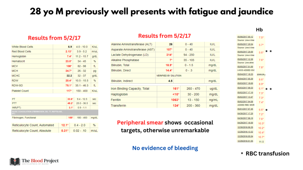
Case
What is the diagnosis in 2 words?

One of 13 slides

Two of 13 slides
Which of the following conditions is associated with macrocytic anemia?
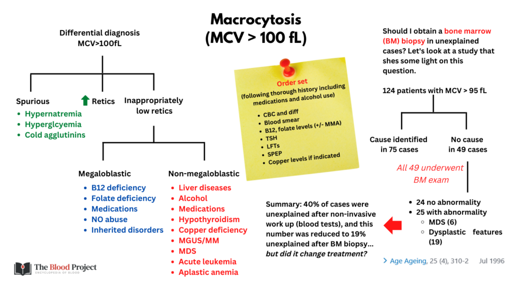
Below is a differential diagnosis for macrocytosis:


Four of 13 slides
Now let’s look at the patient’s CBC over time:


Five of 13 slides
The following were the patient’s reticulocyte counts:


Six of 13 slides
On the first slide it was indicated that the patient had no evidence of bleeding. That leaves hemolysis as the most likely cause.
Seven of 13 slides
The following is a review of commonly ordered hemolytic markers. Note how closely they overlap with changes in liver disease:


Eight of 13 slides
The hemolytic markers in this patient are shown here:


These data are consistent with (more than one answer may apply):
Ten of 13 slides
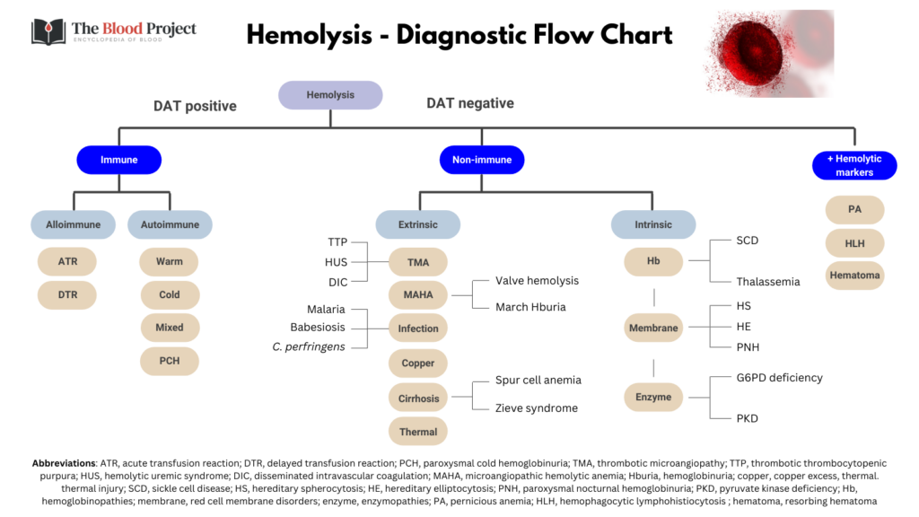
Below is a differential diagnosis for hemolysis:


Eleven of 13 slides
You were told on the first slide that the peripheral smear was unremarkable. That is very helpful in narrow our differential diagnosis of hemolytic anemia:
| Condition | Findings on smear (in addition to polychromatophilia) |
|---|---|
| Immune-mediated | |
| Warm autoimmune hemolytic anemia | Spherocytes |
| Cold autoimmune hemolytic anemia | RBC agglutination |
| Non-immune mediated | |
| Intracorpuscular | |
| Hereditary spherocytosis | Spherocytes |
| Hereditary elliptocytosis | Elliptocytes |
| G6PD deficiency | Normal or with bite cells |
| PK deficiency | Spiculated RBCs resembling spur cells, nRBC |
| SCD | Sickle cells, Howell-Jolly bodies, nRBC |
| Thalassemia | Microcytes |
| PNH | Normal |
| Extracorpuscular | |
| TTP and other TMA | Schistocytes |
| DIC | Normal or with occasional schistocytes |
| Valve hemolysis | Schistocytes |
| Cirrhosis with spur cell anemia | Spur cells (acanthocytes) |
| Zieve syndrome | Normal or with stomatocytes, target cells |
| Wilson disease | Normal |
| Clostridium perfringens | Microspherocytes |
| Babesiosis/malaria | RBC inclusions |
Note that a normal smear in the setting of hemolysis is most consistent with:
- G6PD deficiency
- PNH
- Wilson disease
- Zieve syndrome
Twelve of 13 slides
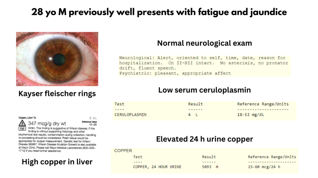
So far, we have a 28 yo M, previously well, presenting with fatigue and jaundice, found to have a hyperproliferative macrocytic anemia, shorted RBC survival consistent with hemolysis, hemolytic markers consistent with both hemolysis and liver disease, and a normal blood smear. Of the diagnoses most consistent with hemolysis + normal peripheral smear (see last slide), only Wilson disease and Zieve syndrome are associated with significant liver disease. It was stated in the initial presentation that that the patient was previously well, which makes Zieve syndrome less likely (as does the extent of liver failure). Indeed, the patient was diagnosed with Wilson disease!
Note that we have approached this case from a hematological angle (for teaching purposes). Of course, when the patient was admitted, he was assessed by the liver service (and hematology, among other services, was consulted). The liver team would have used a diagnostic scoring system (e.g., the Leipzig score) to secure the diagnosis. The Leipzig score includes the following parameter:


Patient results:


| Parameter | Value | Score |
|---|---|---|
| KF rings | Present | 2 |
| Neurological | Normal | 0 |
| Serum ceruloplasmin | 4 mg/dL | 2 |
| Coombs negative HA | Present | 1 |
| Liver copper | 347 mcg/g | 2 |
| Urinary copper | 5093 mcg | 2 |
| Mutation analysis | Not done | 0 |
| Total | 9 |
Easily meets the criteria for diagnosis of Wilson disease.
Thirteen of 13 slides

