Splenomegaly – Differential Diagnosis
Prev
1 / 0 Next
Prev
1 / 0 Next
Splenomegaly is typically defined as an enlarged spleen:
- Weighing > 250 g
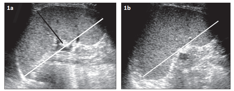
- Width > 4-5 cm, diameter > 7 cm, and length > 11 cm in adults by ultrasound
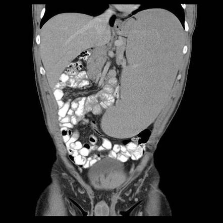
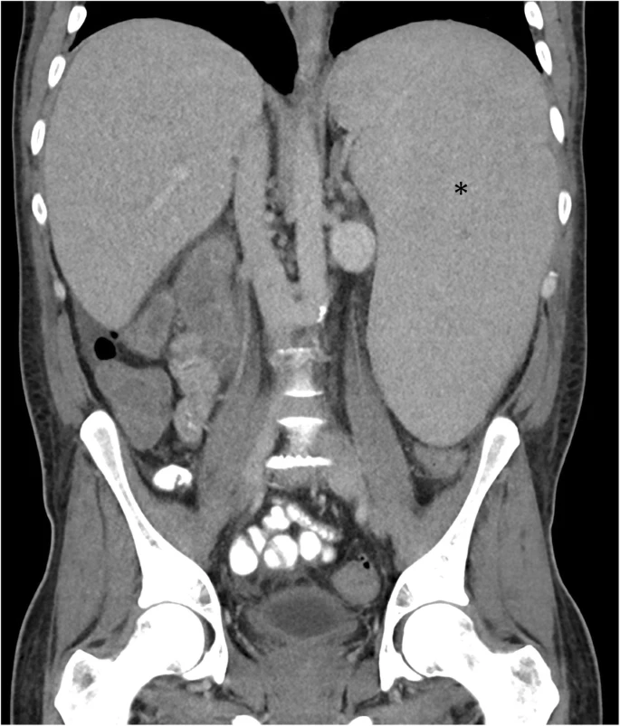
- Maximal craniocaudal dimension of 13 cm by computed tomography (CT)
Enlargement of the spleen occurs through one or more mechanisms (see following slides):
- Work hypertrophy
- Infiltration
- Congestion
Other classification schemes include:
- Massive vs. non-massive splenomegaly
- Isolated splenomegaly or splenomegaly associated with other findings
Detecting splenomegaly:


Splenomegaly may be classified several ways:
- According to prevalence; most common causes include:
- Hematologic (especially hematologic malignancy)
- Hepatic
- Infection
- According to mechanism
- Increased splenic function (work hypertrophy), resulting from excessive function of normal splenic activities such as:
- RBC sequestration (reticuloendothelial system hypertrophy), as seen in:
- Hemoglobinopathies
- Membranopathies
- Immune-mediated hyperplasia, as seen in
- Infections
- Viral
- CMV
- EBV
- Hepatitis
- Bacteria
- Brucellosis
- Tickborne disease
- Tb
- Fungal
- Parasitic
- Visceral leishmaniasis
- Malaria
- Echinococcosis
- Schistosomiasis
- Viral
- Chronic inflammation
- Chronic autoimmune disorders
- ITP
- AIHA
- SLE
- RA
- Felty syndrome
- Sarcoidosis
- HLH
- Hyperthyroidism
- Chronic autoimmune disorders
- Infections
- Extramedullary hematopoiesis, as seen in essential thrombocythemia, polycythemia vera, and primary and secondary myelofibrosis.
- RBC sequestration (reticuloendothelial system hypertrophy), as seen in:
- Infiltration involving abnormal intracellular or extracellular deposition of substances in the spleen secondary to:
- Neoplasms
- Lymphoma
- Leukemia
- ALL
- CML
- HCL
- WM
- Metastatic lesions
- Primary vascular neoplasms of the spleen
- Benign
- Hemangiomas
- Lymphangioma
- Hamartomas
- Littoral cell angiomas
- Malignant – angiosarcoma
- Benign
- Metabolic conditions
- Primary amyloidosis
- Glycogen storage disease
- Rosai-Dorfman disease
- Gaucher disease
- Pseudocysts or true cysts
- Splenic abscesses
- Bacterial
- Parasitic
- Mycotic
- Neoplasms
- Passive congestion due to obstruction of venous blood flow; for example cirrhosis with portal hypertension, heart failure, and splenic/portal/hepatic vein thrombosis.
- Increased splenic function (work hypertrophy), resulting from excessive function of normal splenic activities such as:
- According to whether the splenomegaly is massive (not precisely defined radiologically, but usually defined as clinically palpable > 8 cm below left costal margin or when the lower spleen pole is within the pelvis or when the spleen crosses the midline), seen most commonly in:
- hematological disorders
- Chronic myeloid leukemia
- Agnogenic myeloid metaplasia
- Polycythemia vera
- Essential thrombocythemia
- Indolent lymphomas
- Hairy cell leukemia
- Beta-thalassemia major
- Infectious diseases
- Kala-azar (visceral leishmaniasis)
- Malaria
- infiltrative conditions
- Gaucher disease
- Primary angiosarcoma of the spleen
- hematological disorders
Prev
1 / 0 Next

